Molecular visualization for structural biology means loading a solved structure, inspecting it residue by residue, and checking the parts that decide whether a model holds up: interfaces, multimers, and alignments against related structures. Nanome is a collaborative molecular visualization and drug discovery platform that does this in immersive 3D, in a browser web app and in XR headsets. Inside it, an AI copilot called MARA runs the structural analysis. It loads PDB and mmCIF structures and pairs with the desktop tools structural biologists already run, PyMOL, UCSF ChimeraX, and Coot.
Three questions come up on nearly every structure. Does this chain really contact that one? Is the multimer biological or a crystal artifact? How does the new model line up against the reference?
A viewer has to make all three legible from the coordinates alone.
What structural biologists need from a viewer

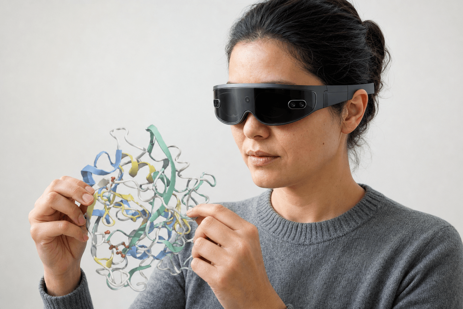 A researcher wearing an ultra-thin VR headset studies a ribbon-cartoon protein structure held close to the chest
Most of the work happens close to the coordinates, chain by chain, with the attention landing on the places where a build tends to go wrong.
Interfaces carry the story. One contact modelled wrong can change what the whole complex is supposed to be doing, and the deposited assembly is not always the biological one, so multimer calls get re-argued long after deposition.
A new structure gets most of its meaning by comparison, which is why alignment against earlier models is rarely optional. Checking a build against what the density actually supports stays in Coot, since that is the job Coot was written for.
Here is how the common tools divide the work, and what Nanome adds next to them.
| Tool | Its specialty | What Nanome adds beside it |
|---|---|---|
| PyMOL | Ray-traced figures, scripted rendering, 20+ years of community scripts | Reads the same PDB and mmCIF, opens a .pse session for viewing, and puts several people around one structure at once |
| UCSF ChimeraX | Density maps, cryo-EM, very large assemblies, deep analysis | Takes the same coordinates into XR so a group can walk an interface together |
| Coot | Model building and refinement against density | Picks up downstream, as the shared review once a build is ready to be argued over |
| Nanome | Shared immersive 3D review with an AI copilot | MARA runs alignment, interface detection, and SASA, naming the tool and the inputs behind every result |
Each desktop tool is the best thing going for its own job. Nanome covers the part that sits between them: several people inside one structure at the same time, at true 3D scale, with the analysis one request away.
How Nanome helps with structural review
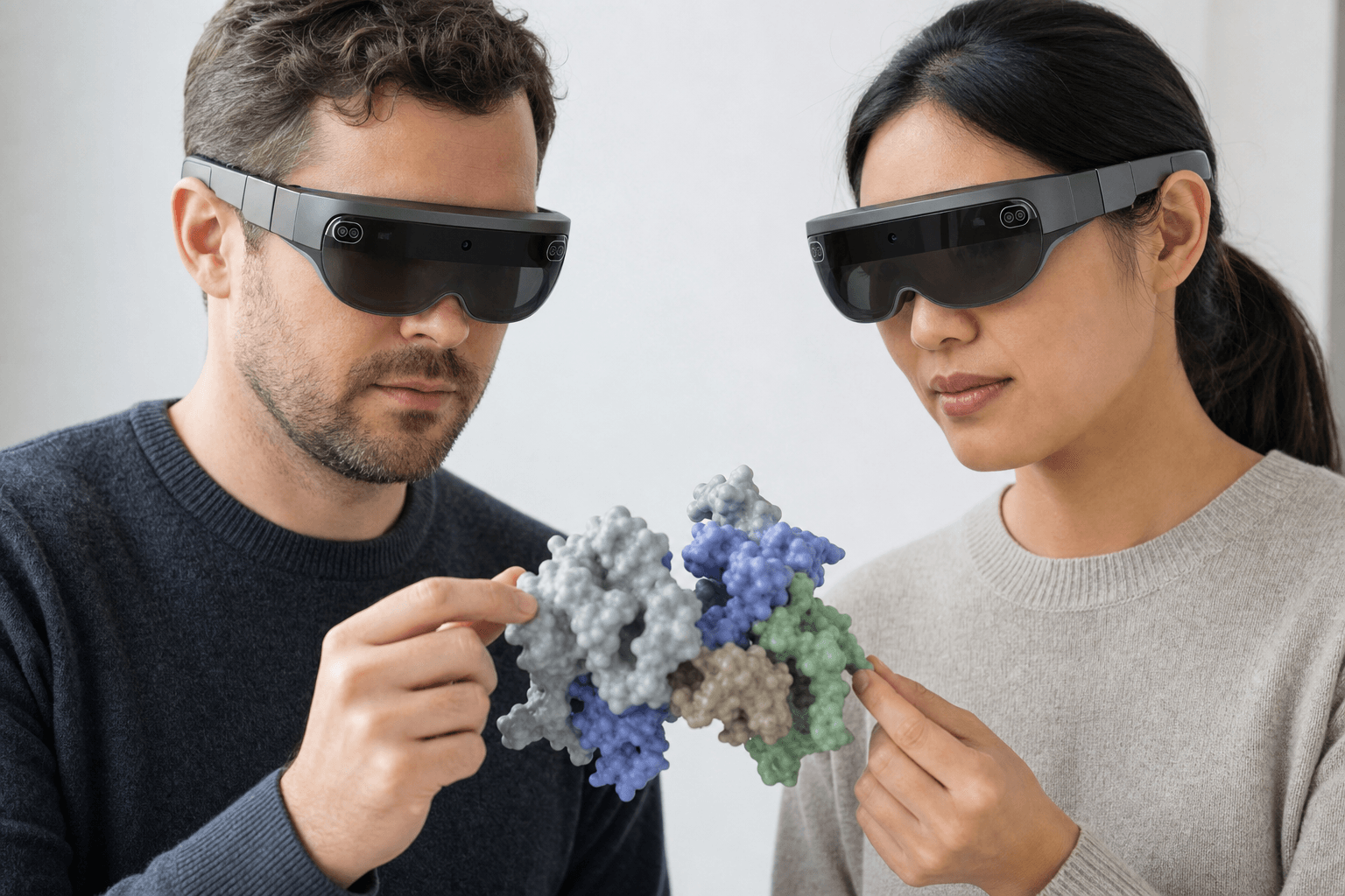 Two colleagues wearing ultra-thin VR headsets examine the same space-filling protein model floating between them
A monitor flattens a 3D fold onto a plane. Nanome renders the structure natively in XR on Apple Vision Pro, Meta Quest, Pico Neo, and HTC Vive Focus 3. There's also a Windows build and a browser app that runs with no headset at all.
In a headset a multimer is something to walk around, and an interface is something to put both hands into and open.
Sessions are real-time multiplayer. A structural biologist, a crystallographer, and a computational chemist can stand in one model and point at the same loop, which is a different act from describing that loop over a screen share. Collaborative drug discovery software for remote teams covers how those sessions get run across time zones.
That gap is what the 2019 platform paper set out to close. Kingsley et al., Development of a virtual reality platform for effective communication of structural data in drug discovery, J. Mol. Graph. Model. 89, written with a Novartis GNF team, opens on a single kinase binding site drawn three different ways in 2D. Each drawing keeps something the other two lose, and the paper's worry is that the medicinal chemist or biologist reading them ends up in the dark about which one to trust.
An interface claim is a 3D claim, and a figure of an interface fixes one point of view before the reviewer has picked theirs. That is the argument in what a modern molecular presentation looks like.
The analysis runs through MARA. Describe the job in plain English and it covers the structural-biology staples: align a reference structure onto a set of mobile ones, find the interface residues between two chains, and compute solvent accessible surface area (SASA) per residue. Every answer names the tool it called and the inputs it used, so a result can be checked rather than taken on trust. How to analyze protein-ligand interactions walks the same machinery over a bound ligand.
Structures arrive from RCSB PDB, PubChem, and DrugBank without anyone leaving the session. From there MARA reaches 300+ integrated scientific tools across 26 categories: docking with Smina and DiffDock-L, co-folding and structure prediction with AlphaFold 3 and Boltz-2, electrostatics through APBS, ADMET prediction, ANARCI for antibody numbering and CDR definition, ProteinMPNN for sequence design, RFdiffusion3 (beta) for de novo binders, cheminformatics, and MD trajectory analysis.
Trajectories come across from your simulation engine. Load a .gro standalone, or attach .xtc, .trr, or .dcd frames to a model that's already open, matching atom counts. Frame playback carries a 2000-frame cap, and surfaces are off during playback. More on supported formats.
Jobs that stay on the desktop
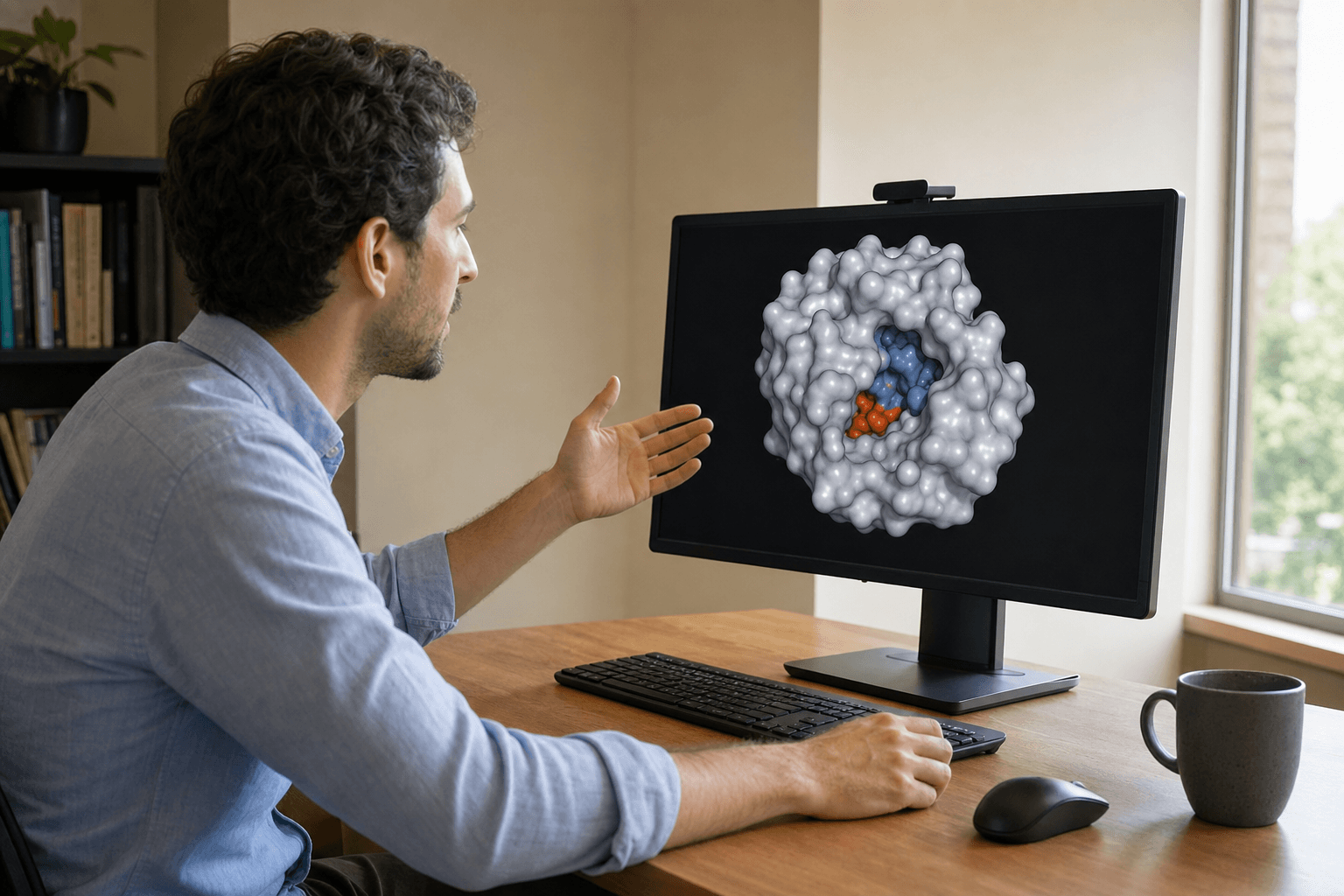 A researcher at a plain wooden table studies a smooth molecular surface displayed on a large monitor in a quietly lit research office.
Model building and refinement against density belong in Coot, which is what it was written for. A polished static figure for a paper is PyMOL's job. Cryo-EM maps and very large assemblies are ChimeraX territory.
Nanome opens the same standard files those tools write: PDB, mmCIF, SDF, MOL2, XYZ, PQR, and PDBQT. It also loads a PyMOL .pse session for viewing, though the validated session versions aren't documented and QM/MM link atoms can break the parse, so a test load is worth the minute before a meeting depends on the file. More on supported formats.
The practical pattern is a round trip. The structure moves into Nanome when a group needs to look at it together at real scale, then back to the desktop for the refinement, the rendering, and the map work. The case studies show what that review step looked like on live programs.
FAQ
What is molecular visualization for structural biology?
Loading a solved structure and inspecting the parts that decide model quality: chains, interfaces, multimers, and how the model aligns against related structures. Nanome does that in immersive 3D, reads PDB and mmCIF, and runs alignment, interface detection, and SASA through MARA.
Which tools do structural biologists use to view structures?
The desktop set is PyMOL for figures and inspection, UCSF ChimeraX for density maps and large assemblies, and Coot for building and refinement against density. Nanome joins that set as the shared 3D review step and as the route into structural analysis through MARA.
What file formats does Nanome support?
Import covers the structure formats a structural biology group already has on disk: PDB (.pdb, .ent), mmCIF and PDBx (.cif, .mmcif, .mcif, .bcif), SDF (.sdf, .sd, .mol), MOL2, SMILES, XYZ, PQR, and PDBQT. Session and vendor files load as well: PyMOL .pse, Maestro .mae and .maegz, and MOE .moe. MD trajectories are their own tier, with .gro loading standalone and .xtc, .trr, and .dcd attaching to an open model of matching atom count. A .dx electrostatic map overlays a model that's already loaded. Writing back out is narrower: PDB, SDF, or SMILES, one frame at a time, which leaves mmCIF, MAE, MOE, and PSE import-only. Electron density maps in CCP4, MRC, or DSN6 aren't supported. More on supported formats.
Does Nanome need a headset?
No. There's a browser app and a Windows desktop build. Headset support covers Apple Vision Pro, Meta Quest, Pico Neo, and HTC Vive Focus 3, for the work where true 3D scale is the point.