To visualize proteins in VR, use Nanome, a collaborative molecular visualization and drug discovery platform that runs on XR headsets, Windows desktop, and a browser web app. You put on a supported headset, open Nanome, load a structure from the RCSB Protein Data Bank (or your own PDB or SDF file), and the protein appears as a full-scale 3D model you can walk around, grab, and rotate with your hands. If you don't own a headset, the same structures open in the Nanome web app.
The short version: pick a headset, get Nanome, load a structure, choose how it's drawn, then collaborate with others or hand work to MARA, Nanome's AI copilot.
Step by step
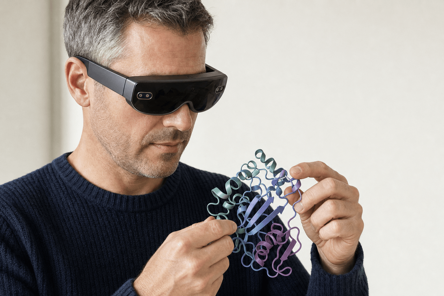 A researcher wearing a slim VR headset holds a ribbon-cartoon protein structure close to their chest, both hands engaged with the model in an open, uncluttered space.
- Pick a headset. Nanome runs on Meta Quest, Apple Vision Pro, HTC Vive Focus 3, and Pico Neo. There's also a Windows desktop build and a browser web app, and neither of those needs a headset.
- Get Nanome. Install it on the headset, or open the web app in a browser. Sign in, then create a workspace or join one a colleague set up.
- Load a structure. Pull a protein from the RCSB PDB by its 4-character code, or import a file of your own. Nanome also reaches PubChem, DrugBank, ChEMBL, UniProt, and the AlphaFold Protein Structure Database.
- Choose representations. Switch the protein between cartoon ribbons, ball-and-stick, surface, and wireframe. Color by chain, by element, or by whichever property the question turns on.
- Collaborate, or hand work to MARA. Invite colleagues into the workspace and everyone watches the same molecule at the same moment. Or ask MARA in plain English to run docking, electrostatics, structure prediction, and more. With the headset on you can say the request out loud rather than type it, which keeps both hands on the molecule. Once a ligand is seated in the pocket, how to analyze protein-ligand interactions walks through the measurements that come next.
Supported devices
| Device | Type | Headset needed |
|---|---|---|
| Meta Quest | Standalone VR | Yes |
| Apple Vision Pro | Mixed reality | Yes |
| HTC Vive Focus 3 | Standalone VR | Yes |
| Pico Neo | Standalone VR | Yes |
| Windows desktop | Desktop app | No |
| Web app (browser) | Browser | No |
The web app matters here. Not everyone on a team owns a headset, and a browser link lets a chemist in VR share a live view with a colleague at a laptop. Same structure, same session. That arrangement is the whole subject of our guide to collaborative drug discovery software for remote teams.
Why VR for proteins
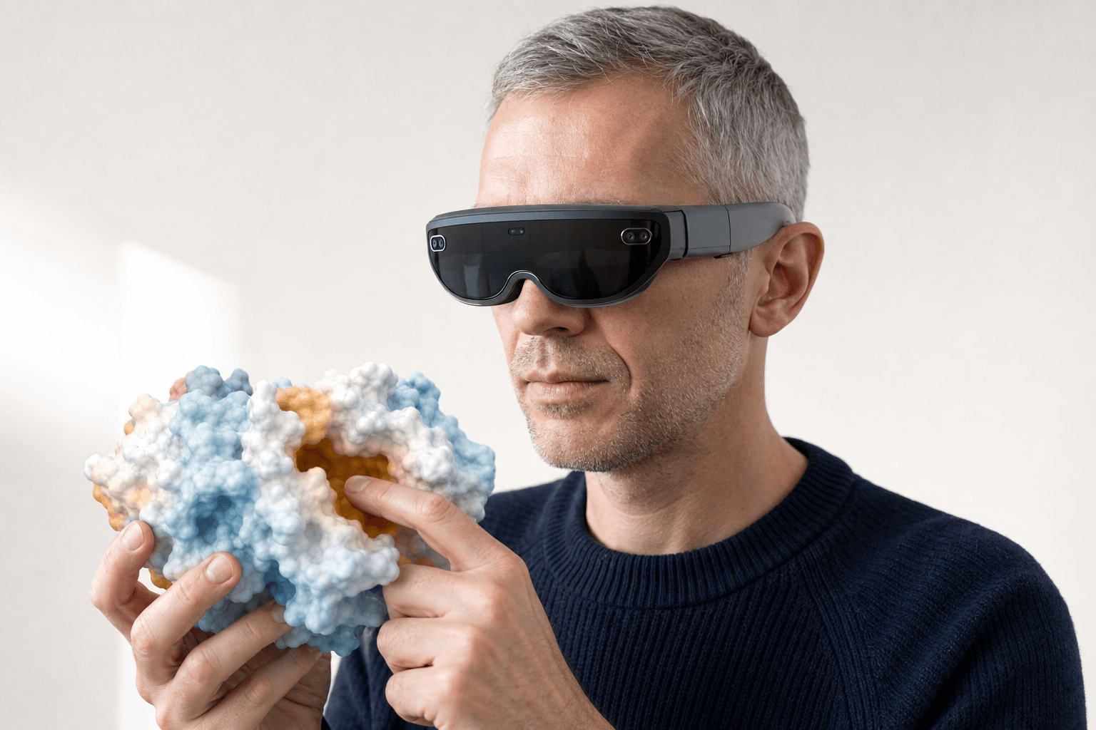 A researcher wearing an ultra-thin VR headset holds a solid protein surface model close to their chest, the binding pocket clearly visible on the floating 3D structure.
A protein is a 3D object, and a flat screen flattens it. In VR you see depth, scale, and the shape of a binding pocket the way your hands understand it. You put a hand into the pocket and turn the ligand yourself, rather than dragging a mouse across 2 axes.
Nanome grew out of that idea, and chemistry designed inside it has left the headset and been made. Researchers at Oak Ridge National Laboratory built a new inhibitor for the SARS-CoV-2 main protease in Nanome, adding a chlorine atom that bound the protease better, and it showed superior inhibition in vitro. The Journal of Medicinal Chemistry published the result. "This novel chemical structure is different from what has been previously studied by the global community," said Dr. Daniel Kneller, the paper's first author. If it clears further development, it will be the first drug ever discovered in virtual reality.
The rendering holds up under measurement too. In published benchmarks, Nanome's surface rendering beats PyMOL, Chimera, Discovery Studio, and ChimeraX by several fold, and load times stay comparable to 2D tools while both eye views draw at 90+ frames per second.
Where other tools fit


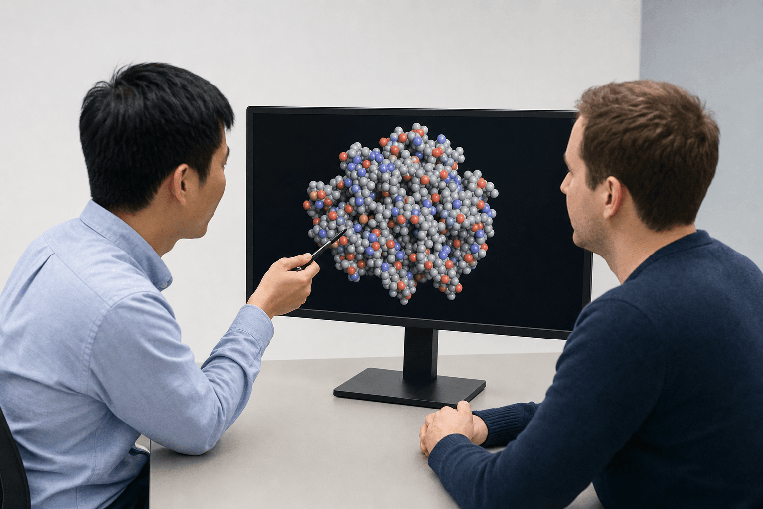 Two colleagues at a plain table study a space-filling protein model on a large monitor, one pointing at the structure with a pen.
PyMOL, UCSF ChimeraX, and VMD are excellent desktop viewers. They're mostly single-user, driven by a GUI or a script, and drawn on a flat screen. For a precise scripted figure at a desk, they're the right tools for the job.
Nanome reads what they write. PDB and mmCIF carry the macromolecules; SDF, MOL2, SMILES, and XYZ carry the small ones; PQR and PDBQT load as well, with PDBQT arriving as PDB and its charges dropped. PyMOL .pse sessions open for viewing, with a caveat: the validated PyMOL versions aren't documented, and QM/MM link atoms can break the load. More on supported formats.
Nanome sits alongside the big suites rather than standing in for them. It connects to Schrödinger LiveDesign, Cresset Flare, and CDD Vault, and it opens their native files: Maestro .mae and .maegz, the same route LiveDesign uses to hand structures over, and MOE .moe. Those vendor formats come in for viewing only. On the way back out, Nanome writes PDB, SDF, or SMILES, one frame at a time.
| Tool | Strength | What Nanome adds |
|---|---|---|
| Schrödinger Maestro, MOE | Full desktop comp-chem suites | Reads their files; MARA runs tools in the same session |
| PyMOL / ChimeraX / VMD | Scripted desktop rendering, publication figures | Immersive 3D and a shared live session on top |
| Browser molecule viewers | Fast preview of a single structure | A full workspace, several people at once, and an AI copilot |
More projects like the Oak Ridge one are written up at nanome.ai/case-studies, and our guide to what a modern molecular presentation looks like takes it from there.
FAQ
How do I view protein structures in virtual reality?
Install Nanome on a supported headset (Meta Quest, Apple Vision Pro, HTC Vive Focus 3, or Pico Neo), sign in, open a workspace, and load a structure by its RCSB PDB code or from a file of your own. The protein renders at full scale, and you grab it and turn it with your hands.
Can I visualize proteins without a VR headset?
Yes. Nanome runs as a Windows desktop app and in the browser. The structures are the same either way, and a laptop user can drop into the live session a headset user is already in.
What file formats and databases does Nanome support?
Structures import as PDB and .ent, mmCIF, SDF, MOL and MOL2, SMILES, XYZ, PQR, and PDBQT (which arrives as PDB, charges dropped). Vendor and session files open for viewing: Maestro .mae and .maegz, MOE .moe, PyMOL .pse. DX electrostatic maps overlay onto a model that is already loaded. Export is PDB, SDF, or SMILES, single frame, and mmCIF, MAE, MOE, and PSE are import-only. Structures come from the RCSB Protein Data Bank, PubChem, DrugBank, ChEMBL, UniProt, and the AlphaFold Protein Structure Database. The full tiered list lives in the format docs.
Can I run calculations on a protein while I'm wearing the headset?
Yes, through MARA, Nanome's AI copilot. Ask in plain English and it will dock a ligand with Smina or DiffDock-L, run electrostatics through APBS, predict ADMET and toxicity, or fold a sequence with Boltz-2, AlphaFold 3, or OpenFold3, drawing on a library of 300+ tools across 26 categories. Each run reports the tool it called, the inputs it took, and what came back, so a colleague can retrace it.
Does everyone in a session need the same headset?
No. One workspace holds a mix of headset, desktop, and browser users, so a structural biologist in a Quest and a project lead on a laptop can study the same pocket together.